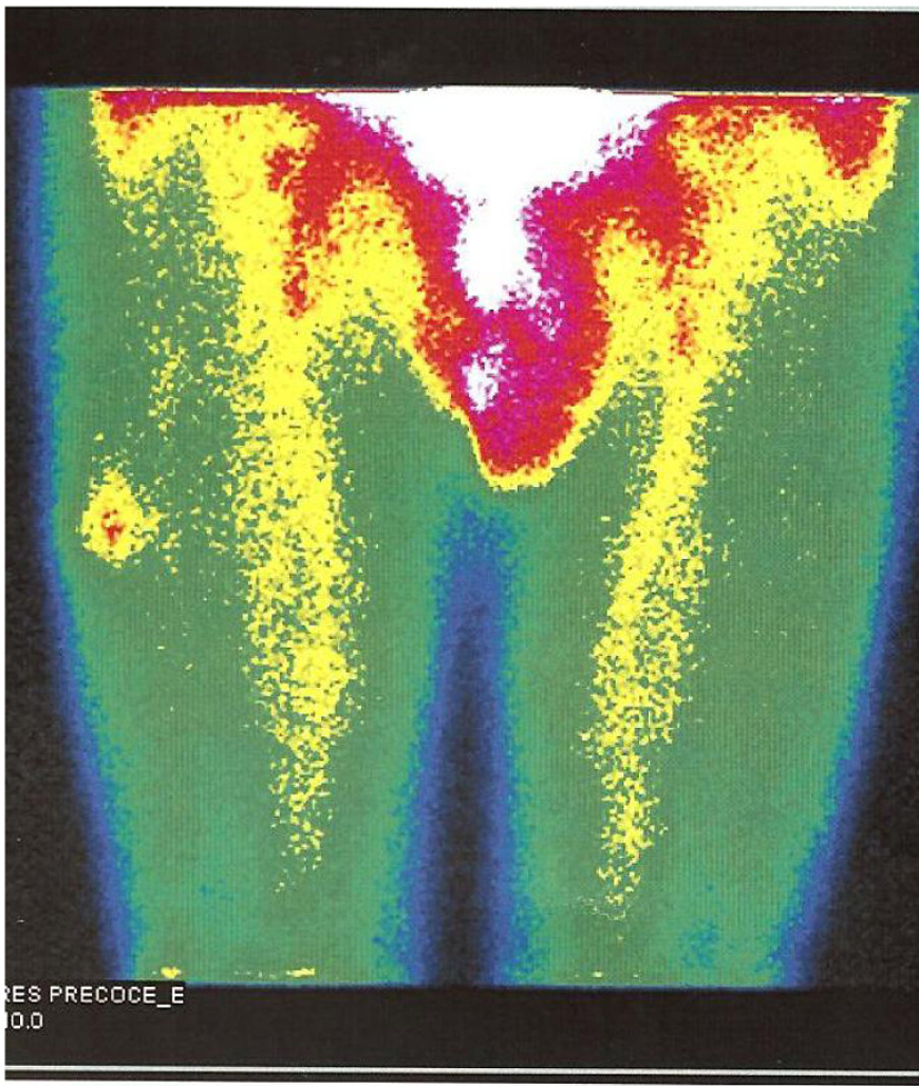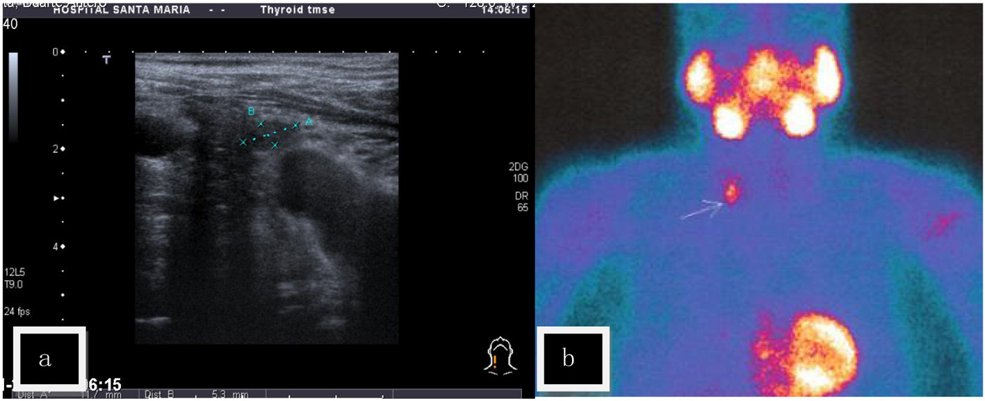
Figure 1. (a) Cervical sonography (US) - a nodular lesion was found contiguous to the lower right pole of the thyroid gland. (b) Sestamibi scintigraphy - positive late fixation at the right lower pole of the thyroid.
| Journal of Endocrinology and Metabolism, ISSN 1923-2861 print, 1923-287X online, Open Access |
| Article copyright, the authors; Journal compilation copyright, J Endocrinol Metab and Elmer Press Inc |
| Journal website http://www.jofem.org |
Case Report
Volume 5, Number 1-2, April 2015, pages 184-188
Soft Tissue Metastasis of Parathyroid Carcinoma: Description of a Difficult Case
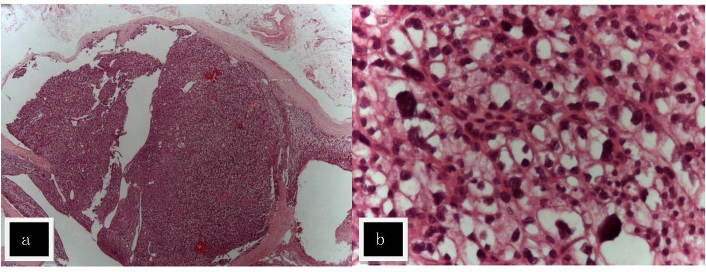
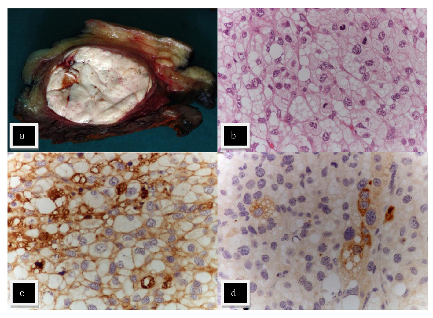
Figures