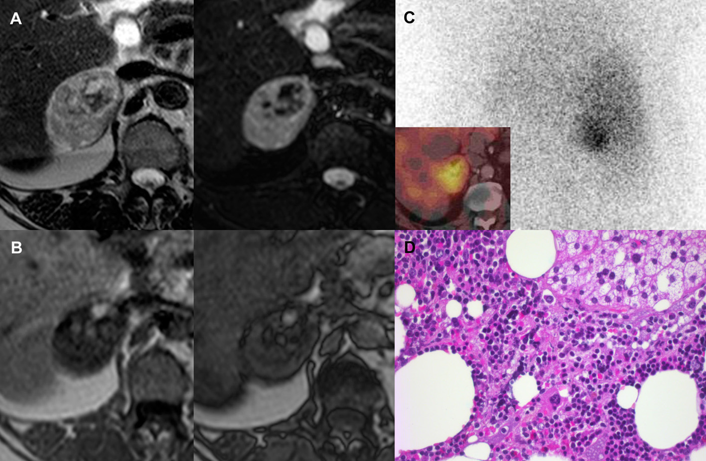
Figure 1. (A) Heterogenous signal on MRI. Fatty content areas with high T2 signal (upper left) and low T2 fat sat signal (upper right). (B) Absence of loss of signal intensity on out-of-phase T1 images (right image) compared to in-phase T1 images (left image), suggesting that there was no intracellular fat. (C) Unilateral uptake visible on 131I-19-iodocholesterol (planar static image, posterior view, SPECT and CT fusion image: small image) which was consistent with a cortisol-secreting adrenocortical tumor. (D) Islands of adrenal tumoral cells admixed with fatty and fibrous territories as well as hemorrhagic areas. Higher magnification showing details of the myeloid tissue with characteristic megakaryocytes (hematoxylin-eosin stain × 400).