Journal of Endocrinology and Metabolism, ISSN 1923-2861 print, 1923-287X online, Open Access
Article copyright, the authors; Journal compilation copyright, J Endocrinol Metab and Elmer Press Inc
Journal website http://www.jofem.org

Case Report

Volume 5, Number 4, August 2015, pages 267-271


18F-Choline PET/CT as a New Tool for Functional Imaging of Non-Proliferating Secreting Neuroendocrine Tumors

Figures

Figure 1.
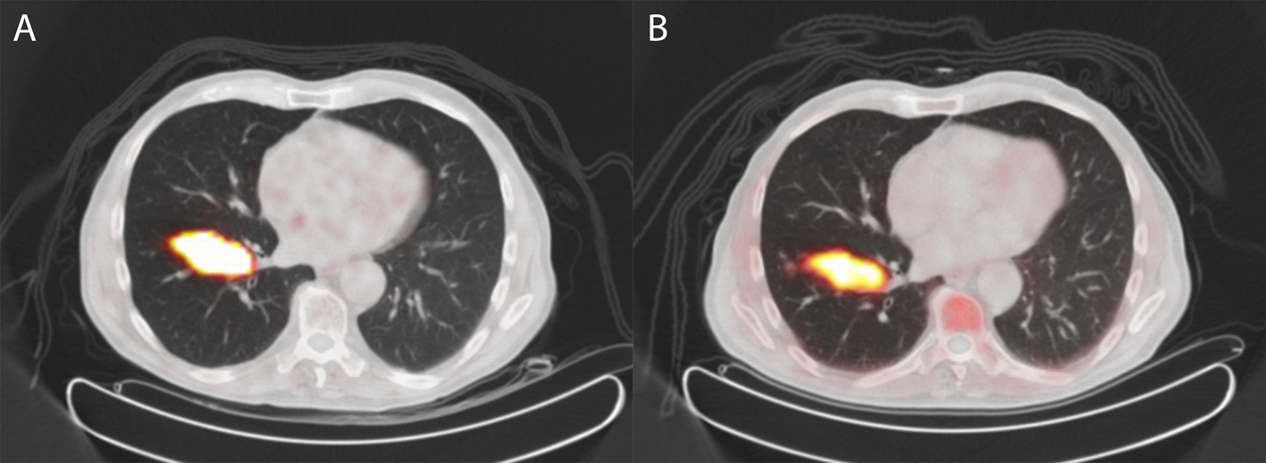
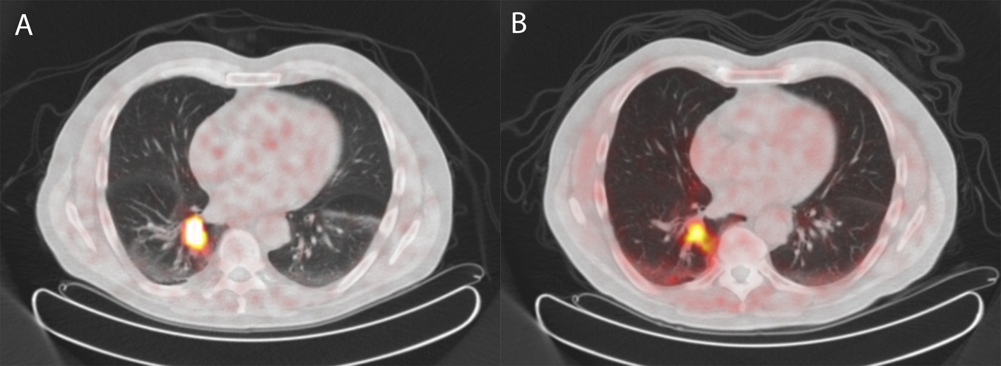
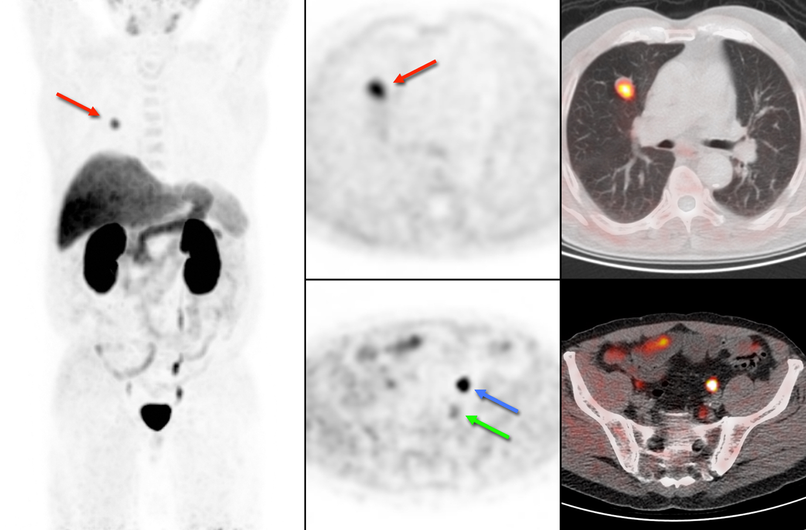
Figure 1. 18F-choline PET/CT of case 1. Physiological uptake of 18F-choline is seen in the salivary glands, liver, spleen and kidneys. The blue arrow shows excretion of radioactive urine in the left ureter and the green arrow indicates pathological uptake in a lymph node. In addition, we encountered an unexpected lung lesion in the right upper lobe with high uptake of 18F-choline (red arrow).
Figure 2.
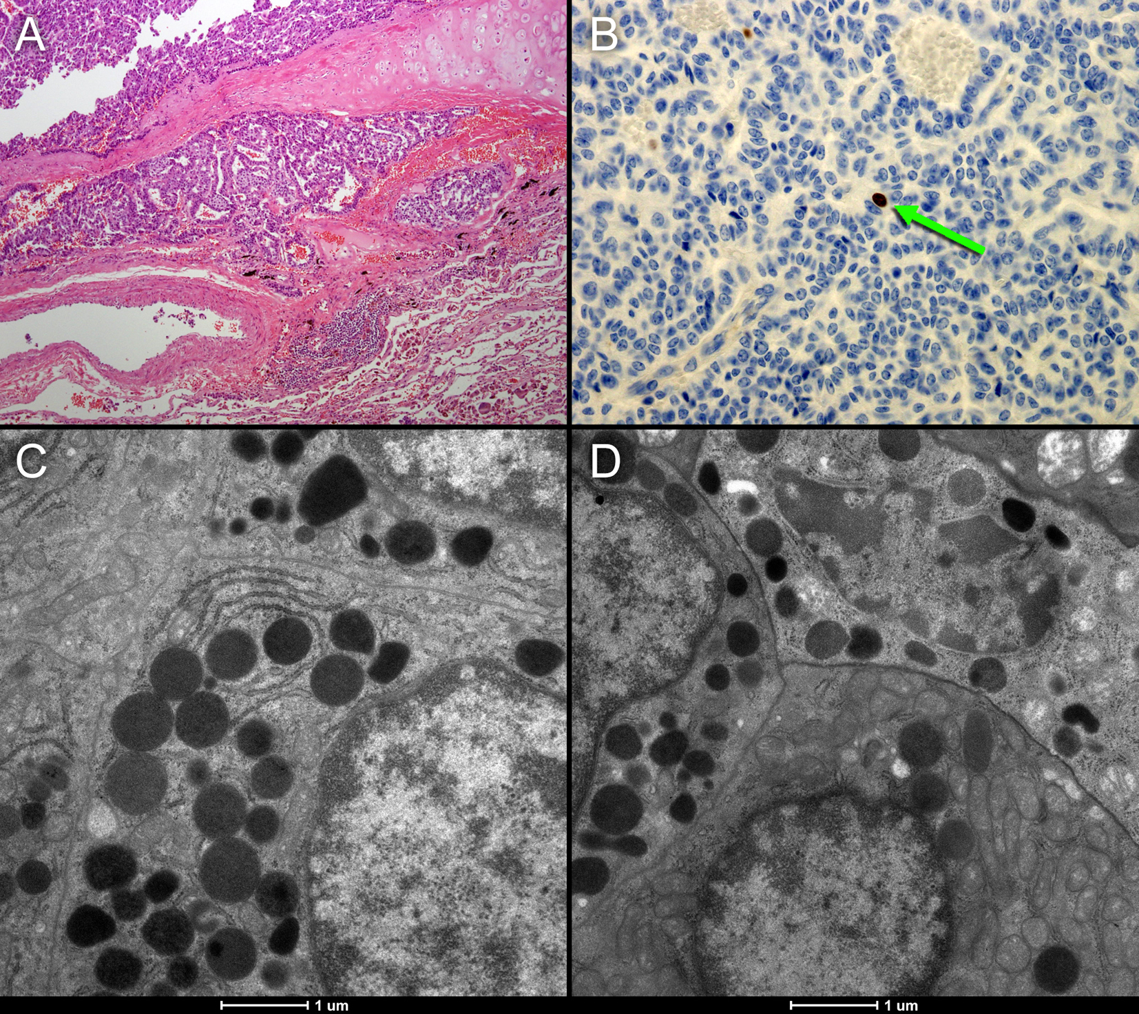
Figure 2. (A, B) Histopathological sample of the resected neuroendocrine tumor of case 1. The tumor shows good vascularization and hardly any necrosis (A). MIB1 immunostaining representing Ki-69 mitotic activity (B: brown colored cells) shows that there is hardly any mitotic activity (< 2 per 2 mm2). The electron microscope images (C and D) of the neuroendocrine tumor show numerous voluminous electron dense vesicles in the cytoplasm (black circles).
Figure 3.
Figure 3. 68Gallium-DOTATATE PET/CT (A) and 18F-choline PET/CT (B) of a patient with an atypical carcinoid of the lung (case 2). The tumor shows increased somatostatin receptor expression on 68Gallium-DOTATATE PET/CT but also intense uptake of 18F-choline on 18F-choline PET/CT.
Figure 4.
Figure 4. 68Gallium-DOTATATE PET/CT (A) and 18F-choline PET/CT (B) of a patient with an atypical carcinoid of the lung (case 3) revealing similar uptake of 68Gallium-DOTATATE as well as 18F-choline as compared to case 2.