Journal of Endocrinology and Metabolism, ISSN 1923-2861 print, 1923-287X online, Open Access
Article copyright, the authors; Journal compilation copyright, J Endocrinol Metab and Elmer Press Inc
Journal website http://www.jofem.org

Case Report

Volume 6, Number 1, February 2016, pages 27-29


A Large Superior Mediastinal Mass: “Terrible Thyroid Cancer”

Figures

Figure 1.
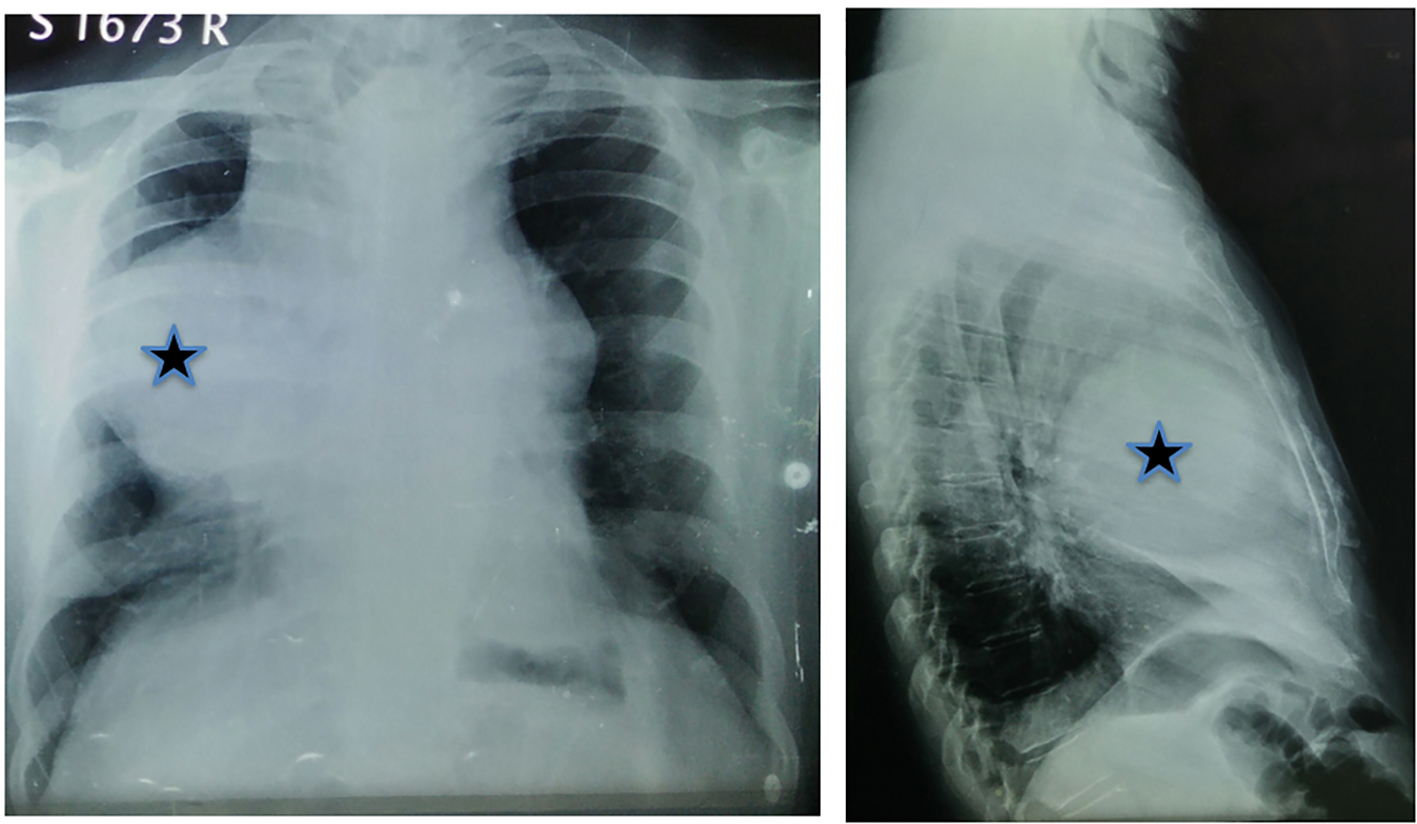
Figure 1. Chest X-ray showing large antero-superior mediastinal mass (star).
Figure 2.
Figure 2. Coronal view FDG-PET-CT: large mediastinal cystic mass with FDG uptake is seen in the periphery of the mass. Also seen is intense FDG uptake on the left lobe thyroid nodule (star) and left level IV cervical lymph node (arrow head).
Figure 3.
Figure 3. Histopathology (HPE) of mediastinal mass showing cyst wall containing hemorrhage and cholesterol clefts (arrow) with periphery showing the lymphoid follicles with black anthracotic pigment (arrow head). The inset (× 20) shows a focus of long papillae lined by single layer of tall columnar cells with sharply delineated cell borders, intensely eosinophilic, finely granular cytoplasm consistent with tall cell variant of PTC.