Journal of Endocrinology and Metabolism, ISSN 1923-2861 print, 1923-287X online, Open Access
Article copyright, the authors; Journal compilation copyright, J Endocrinol Metab and Elmer Press Inc
Journal website http://www.jofem.org

Review

Volume 6, Number 1, February 2016, pages 1-11


Therapeutic Methods Against Insulin Resistance

Figures

Figure 1.
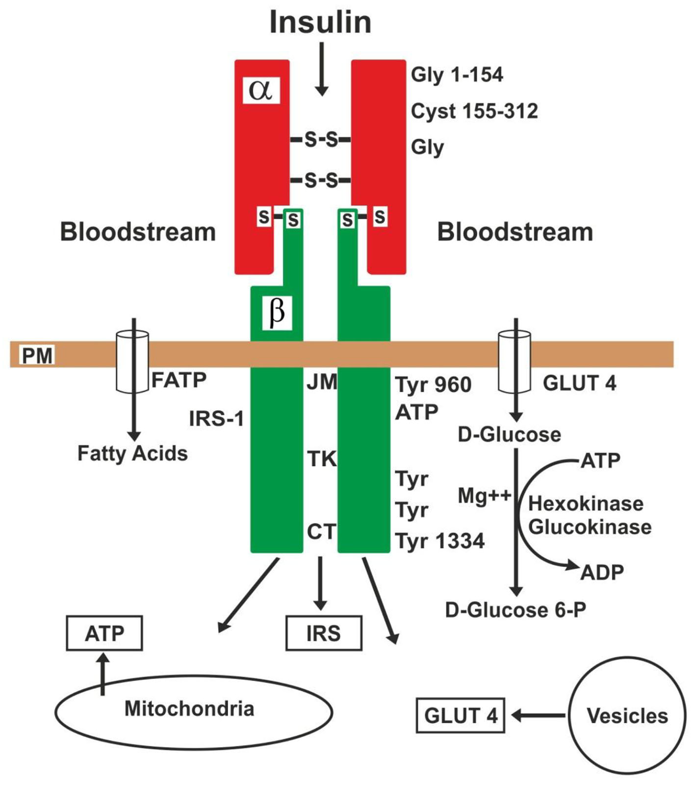
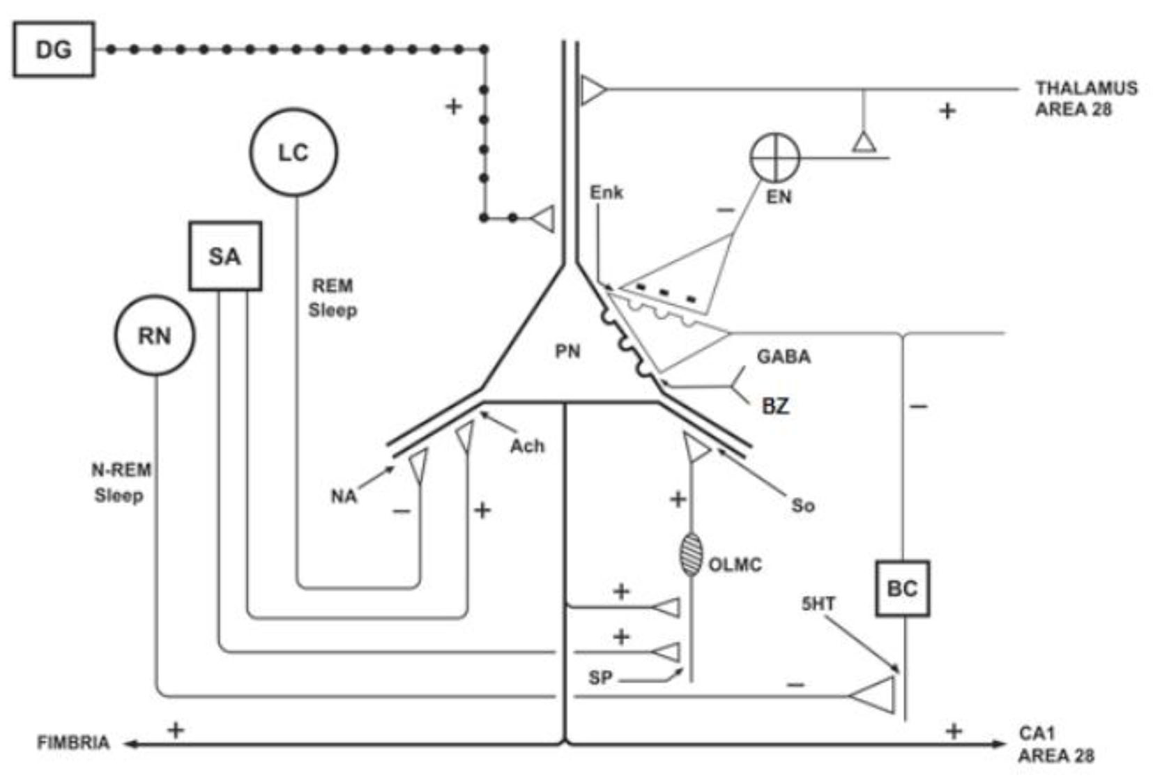
Figure 1. Schematic representation of the hippocampus, showing the ascending projections originating from the septal area (SA), locus coeruleus (LC) and raphe nuclei (RN), and the action of the benzodiazepines (BZ) on the pyramidal neurons (PNs). DG: dentate gyrus; BC: basket cell; EN: enkephalinergic neuron; Enk: encephalin; OLMC: olm cell; SO: somatostatin; Ach: acetylcholine; NA: noradrenaline; 5HT: serotonin; SP: substance P. Adapted from reference [22].
Figure 2.
Figure 2. Action of insulin on its receptor and substrates. PM: plasma membrane; JM: juxtamembrane; TK: tyrosine kinase; CT: carboxyl-terminal: GLUT4: glucose transporter-4; FATP: fatty acid transporter; IRS: insulin receptor substrate; Gly: glycine; Cyst: cysteine; Tyr: tyrosine.
Figure 3.
Figure 3. The insulin receptor is injured by inflammatory cytokines, which cause insulin resistance. On the contrary, this injury can be aborted or improved by loss of body fat and the use of anti-inflammatory drugs.