Figure 1.
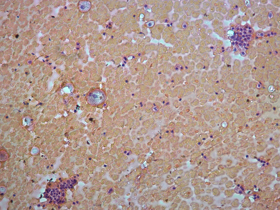
Cytology from thyroid fine needle aspiration. This shows benign follicular cells with increased lymphocytes in the background and scattered macrophages (× 400).
| Journal of Endocrinology and Metabolism, ISSN 1923-2861 print, 1923-287X online, Open Access |
| Article copyright, the authors; Journal compilation copyright, J Endocrinol Metab and Elmer Press Inc |
| Journal website http://www.jofem.org |
Case Report
Volume 7, Number 6, December 2017, pages 190-193
Metastatic Neuroendocrine Tumor in the Thyroid: Report of an Incidental Diagnosis
Figures
Cytology from thyroid fine needle aspiration. This shows benign follicular cells with increased lymphocytes in the background and scattered macrophages (× 400).
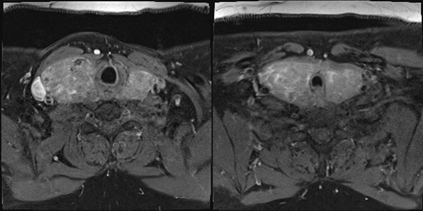
MRI of face and neck. Bilaterally enlarged thyroid lobes, right more than left, laterally deviating carotid arteries and jugular veins.
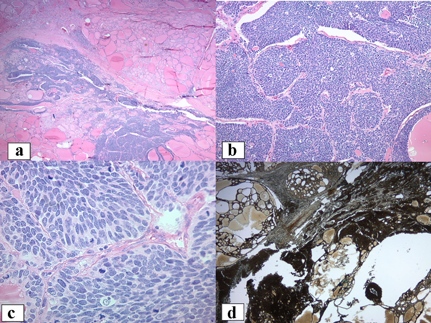
Histologic features of the thyroid. (a, b) These images are in medium power view showing solid sheets and nests of carcinoma in a background of a benign multinodular goiter (× 100, × 200). (c) The tumor cells contain clear to eosinophilic cytoplasm and oval nuclei with fine dispersed, granular chromatin typical of neuroendocrine tumors. Nuclear molding as well as numerous mitoses is present, including atypical forms (× 400). (d) CD-56 immunohistochemistry shows diffuse and strong cytoplasmic staining.
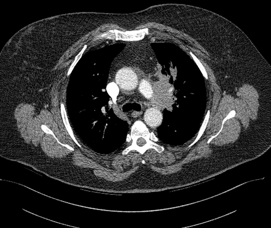
Chest CT. A 68 × 37 × 51 mm soft tissue mass is seen within the mediastinum pre vascular space and exhibits mass effect.