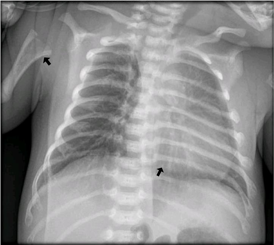
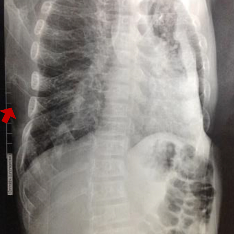
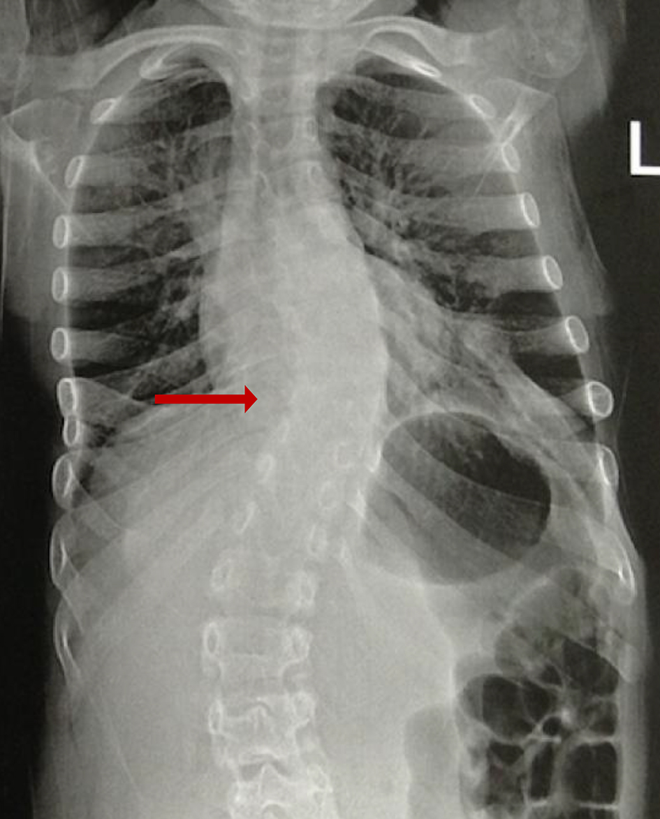
Figure 1. Horizontalization of the rib arches, decreased bone mineral density and slight thickening of the chondrocostal junctions (arrows).
| Journal of Endocrinology and Metabolism, ISSN 1923-2861 print, 1923-287X online, Open Access |
| Article copyright, the authors; Journal compilation copyright, J Endocrinol Metab and Elmer Press Inc |
| Journal website https://www.jofem.org |
Case Report
Volume 11, Number 2, April 2021, pages 56-64
Clinical Variability of Hypophosphatasia in Colombian Patients: Case Reports
Figures
Tables
| Laboratory test | Case 1 | Case 2 | Case 3 | Case 4 | Case 5 | Case 6 | Case 7 |
|---|---|---|---|---|---|---|---|
| ND: no data. | |||||||
| Alkaline phosphatase | < 20 U/L | 9.0 U/L | 116.6 U/L | 114 U/L | 31 U/L | 30 U/L | 27.4 U/L |
| Calcium | 9.5 mg/dL | 9.4 mg/dL | 10.71 mg/dL | 9.3 mg/dL | 9.8 mg/dL | 10.3 mg/dL | 10 mg/dL |
| Phosphorus | 8.6 mg/dL | 4.8 mg/dL | 5.24 mg/dL | 5.75 mg/dL | 3.1 mg/dL | 5.9 mg/dL | 6.4 mg/dL |
| Parathyroid hormone | 19.2 pg/mL | 57.6 pg/mL | 24.0 pg/mL | 26.35 pg/mL | 31 pg/mL | 67 pg/mL | 40.9 pg/mL |
| Vitamin D | 22.7 ng/mL | 17.8 ng/mL | 53.2 ng/mL | 36.4 ng/mL | 38.9 ng/mL | ND | 28.3 ng/mL |
| Case 1 | Case 2 | Case 3 | Case 4 | Case 5 | Case 6 | Case 7 | |
|---|---|---|---|---|---|---|---|
| +: present; -: absent. | |||||||
| Gender | M | M | M | M | M | M | F |
| Age of onset of symptoms | In utero | 3 years | 10 months | 18 months | 2 years | 10 months | 8 months |
| Age of diagnosis | At birth | 3 years | 5 years | 12 years | 13 years | 6 years | 9 years |
| History of motor development delay | - | - | - | + | + | + | + |
| Dental manifestations | NA | + | + | + | + | + | + |
| Fractures | - | + | - | - | - | - | |
| Seizures | - | - | - | - | - | + | - |
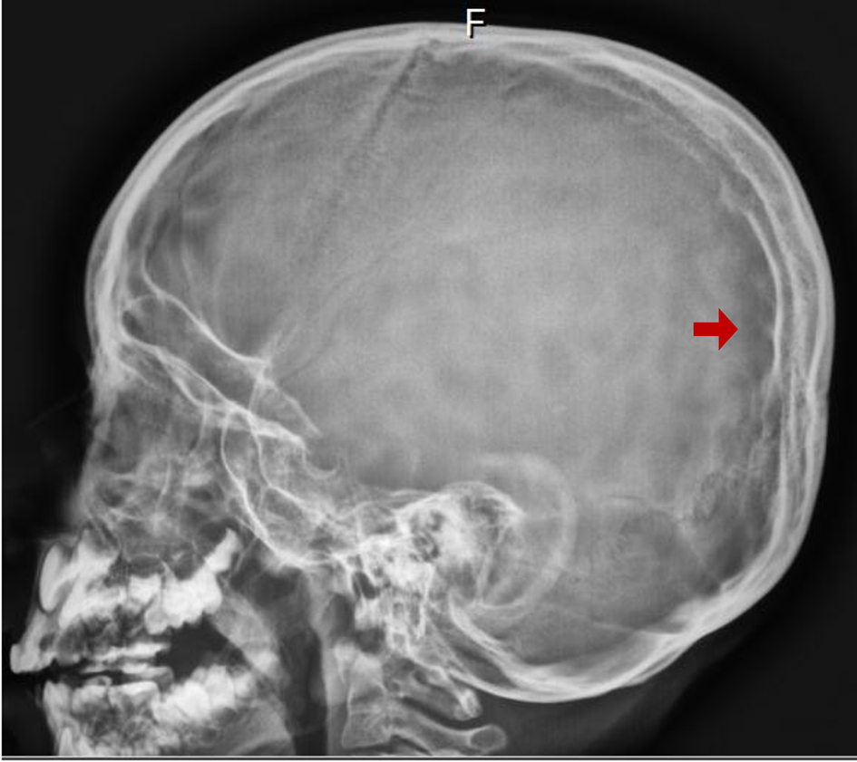
| Craniosynostosis | - | - | + | - | - | + | - |
| Short stature | + | + | + | + | + | + | + |
| Scoliosis | - | + | - | - | - | + | + |
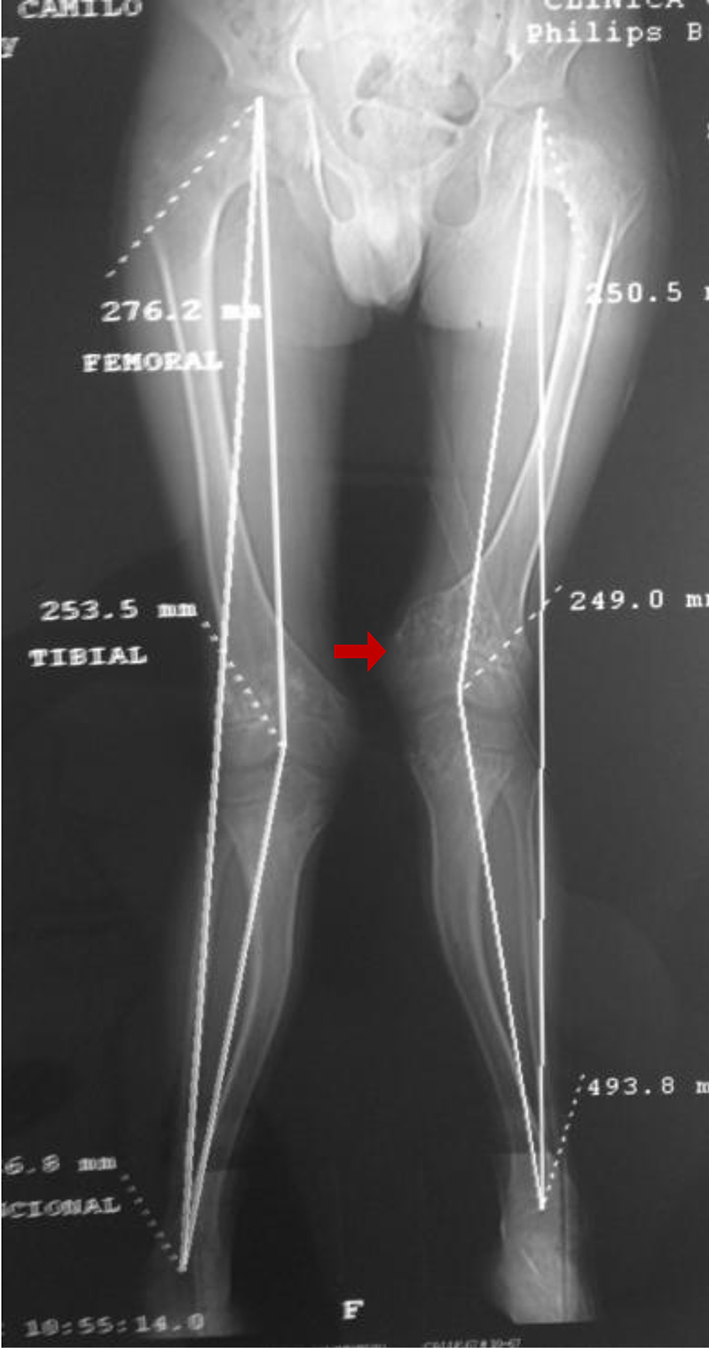
| Limb deformity | + | + | - | + | + | + | + |
| ALPL mutations | p.Glu298Lys/p.Glu298Lys | p.Ala132Thr/p.Glu191Lys | p.Glu298Lys | p.Gly112Arg | p.Glu298Lys/p.Glu298Lys | p.Glu298Lys/p.Glu298Lys | p.Glu298Lys/p.Glu298Lys |
| Type of hypophosphatasia | Perinatal benign | Childhood | Childhood | Childhood | Childhood | Childhood | Childhood |