Journal of Endocrinology and Metabolism, ISSN 1923-2861 print, 1923-287X online, Open Access
Article copyright, the authors; Journal compilation copyright, J Endocrinol Metab and Elmer Press Inc
Journal website https://www.jofem.org

Case Report

Volume 11, Number 2, April 2021, pages 52-55


An Unusual Complication of Cabergoline Treatment of Macroprolactinomas

Figures

Figure 1.
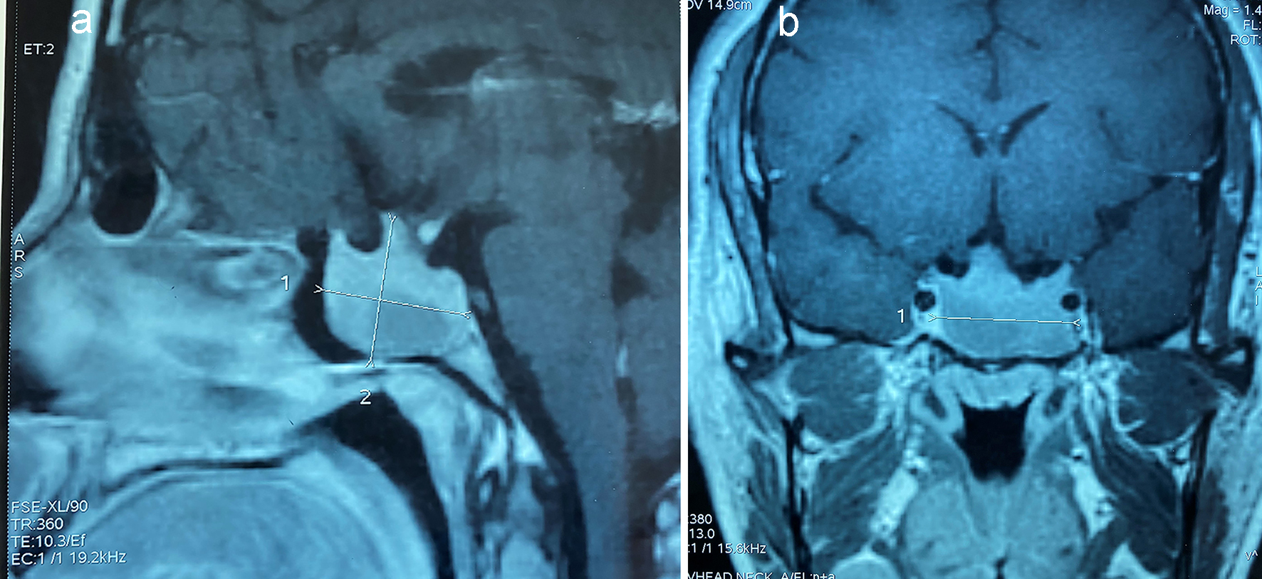
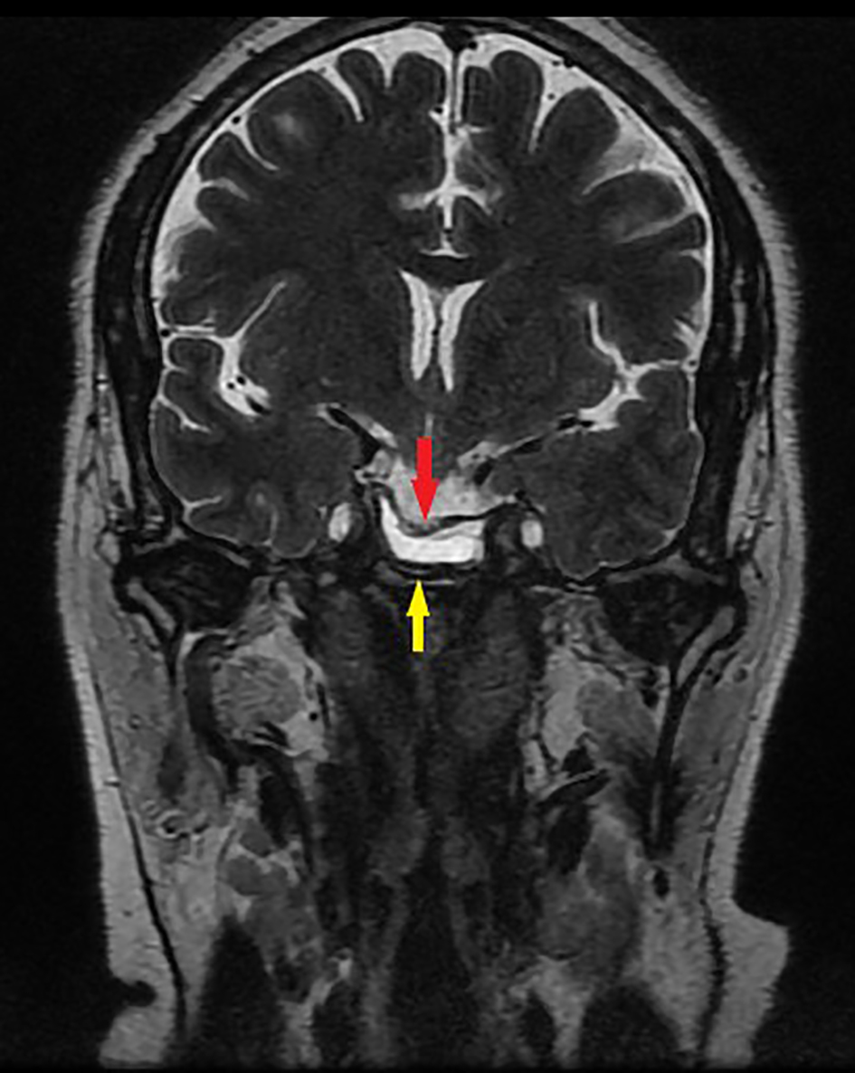
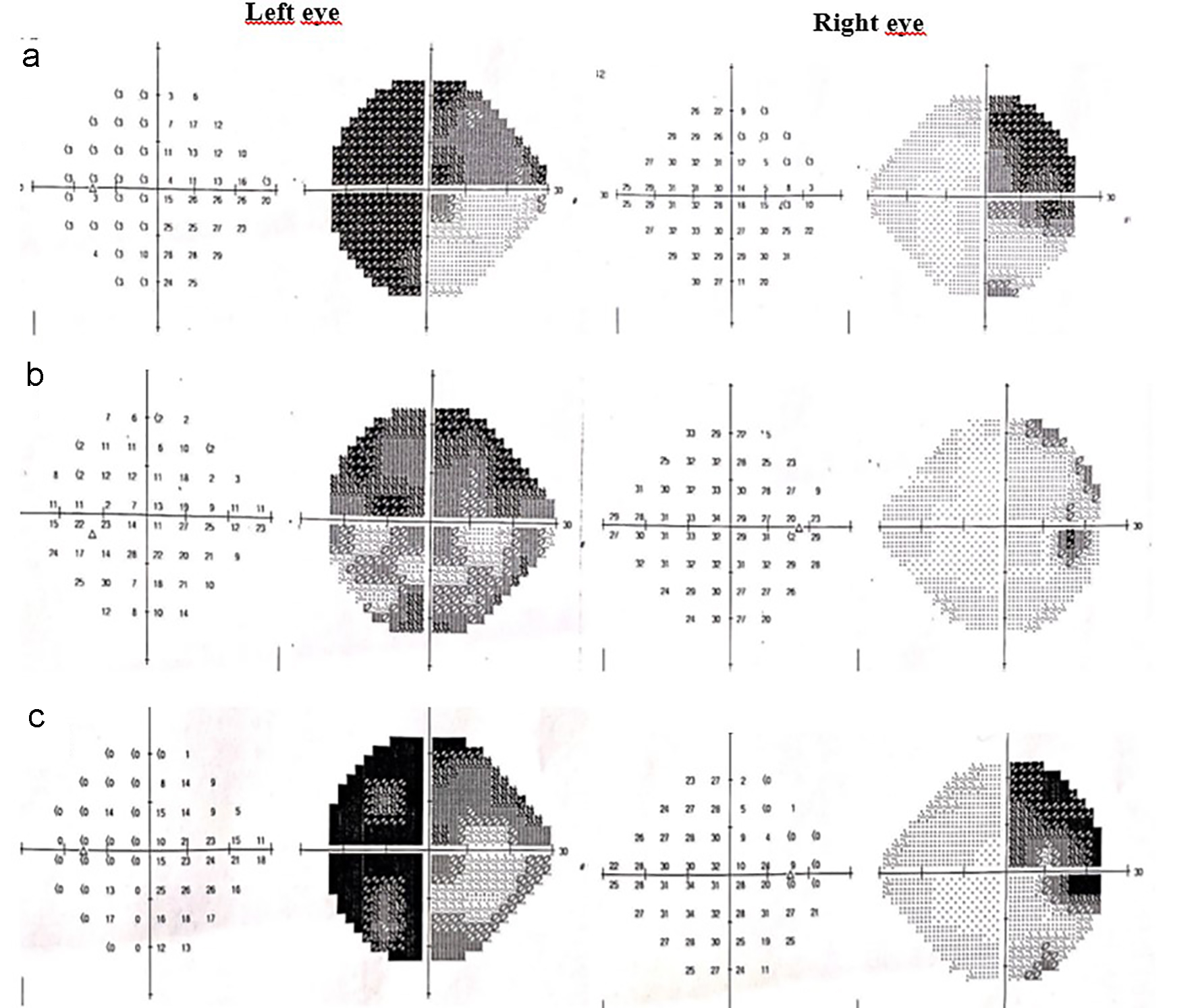
Figure 1. (a) Initial visual fields showing right superior temporal quadrantanopsia and left temporal hemianopsia. (b) After 2 months of cabergoline treatment, visual fields showing improvement in the temporal fields. (c) After 14 months of cabergoline treatment, visual field showing a secondary deterioration of temporal field.
Figure 2.
Figure 2. Sagittal (a) and coronal (b) T1-weighted magnetic resonance image showing a decrease of the adenoma size after 3 months of cabergoline treatment.
Figure 3.
Figure 3. Coronal T2-weighted magnetic resonance image showing shrinkage of the pituitary adenoma (yellow arrow) with prolapse of optic chiasm (red arrow).