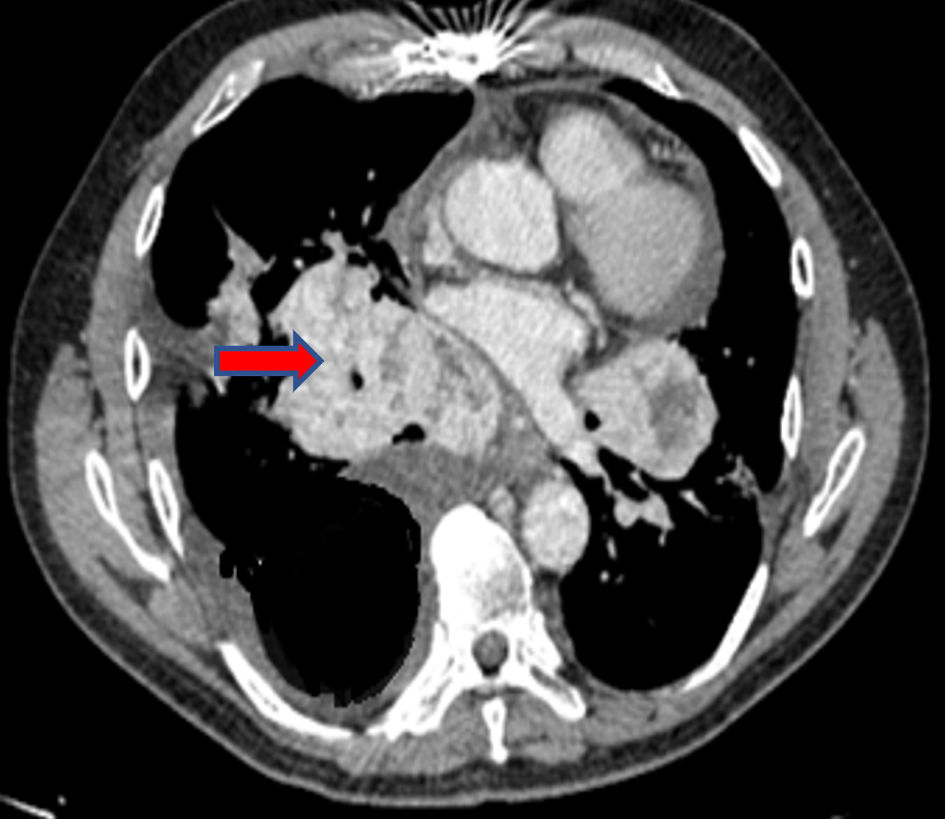
Figure 1. Chest CT scan: expansive lesion in the right anterior mediastinum (arrow). CT: computed tomography.
| Journal of Endocrinology and Metabolism, ISSN 1923-2861 print, 1923-287X online, Open Access |
| Article copyright, the authors; Journal compilation copyright, J Endocrinol Metab and Elmer Press Inc |
| Journal website https://www.jofem.org |
Case Report
Volume 12, Number 6, December 2022, pages 198-201
Mediastinal Mass as First Clinical Manifestation of Medullary Thyroid Microcarcinoma in a Patient With Hashimoto’s Thyroiditis
Figures